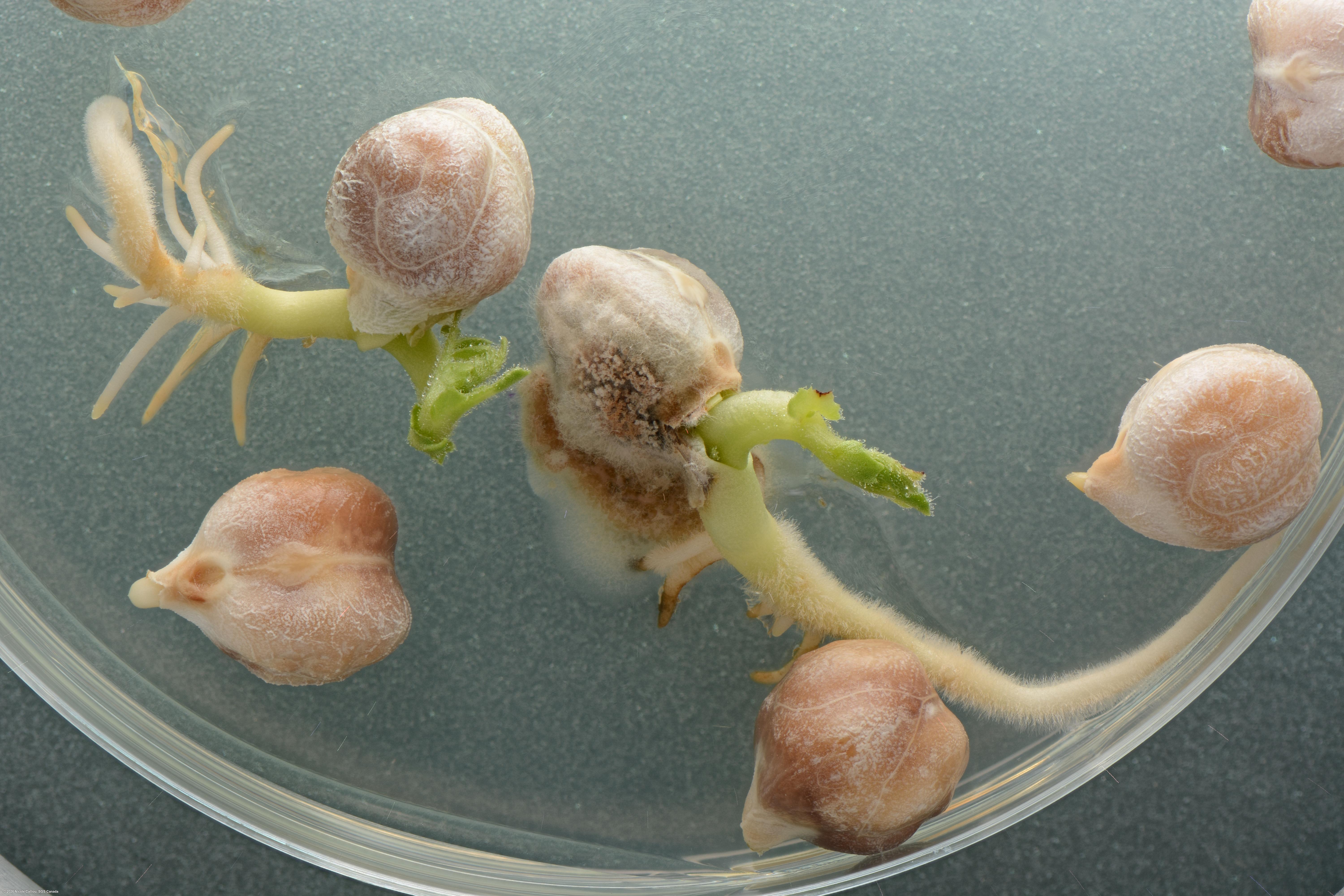
Ascochyta rabiei on Cicer arietinum
Image credits: Nicole Calliou, CA
On PDA, OR on MA, white to green colored mycelium develops slowly, development of pale brown to dark brown pycnidia occurs on surface or immersed. At 7 days, colony diameter varies between 10-35 mm on both media. Pycnidia usually release orange mucilaginous mass of conidia (cirrus). Doubtful colonies may be confirmed by the presence of pycnidia on seed coat or on media surface when examined at x25 magnification, using stereomicroscope. Pycnidia are 65-245 μm diameter, ostiole 30-50 μm wide. Further confirmation can be made by examining conidia present in pycnidia using compound microscope (x100-1000 magnification). Conidia are hyaline, straight, or slightly curved (0-)1 septate, some unicellular, slightly, or not constricted at the septum, rounded at each end, mostly 7-16 x 3-5 μm.
sterilisation: 1% NaOCl 4 minutes | growth time: 7 days | temperature: 22 ± 2 °C | lighting: Alternating 12 hours light and 12 hours dark | plate size: 90 mm | agar: PDA ( Neogen) |
DNA testing: N/A
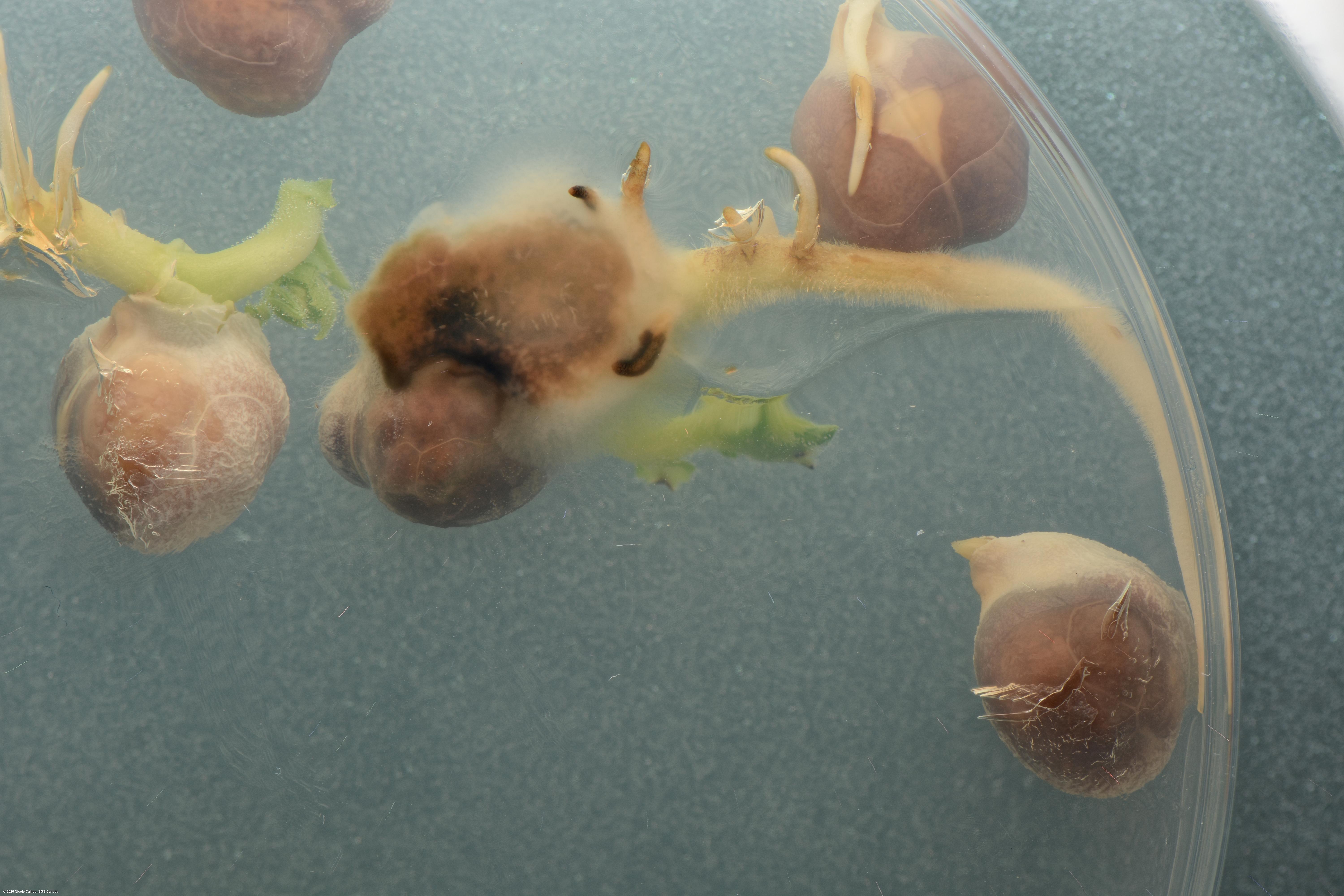
Ascochyta rabiei on Cicer arietinum
Image credits: Nicole Calliou, CA
On PDA, OR on MA, white to green colored mycelium develops slowly, development of pale brown to dark brown pycnidia occurs on surface or immersed. At 7 days, colony diameter varies between 10-35 mm on both media. Pycnidia usually release orange mucilaginous mass of conidia (cirrus). Doubtful colonies may be confirmed by the presence of pycnidia on seed coat or on media surface when examined at x25 magnification, using stereomicroscope. Pycnidia are 65-245 μm diameter, ostiole 30-50 μm wide. Further confirmation can be made by examining conidia present in pycnidia using compound microscope (x100-1000 magnification). Conidia are hyaline, straight, or slightly curved (0-)1 septate, some unicellular, slightly, or not constricted at the septum, rounded at each end, mostly 7-16 x 3-5 μm.
sterilisation: 1% NaOCl 4 minutes | growth time: 7 days | temperature: 22 ± 2 °C | lighting: Alternating 12 hours light and 12 hours dark | plate size: 90 mm | agar: PDA ( Neogen) |
DNA testing: N/A
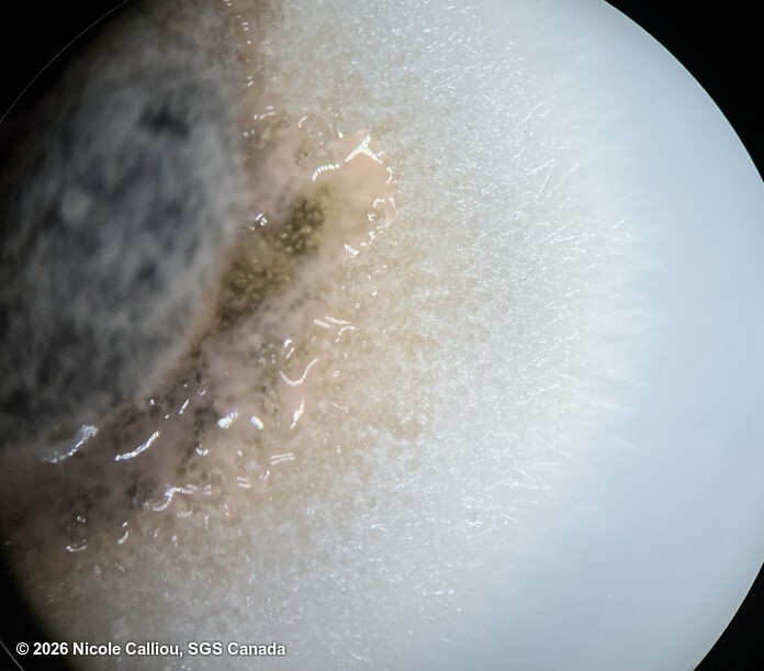
Ascochyta rabiei on Cicer arietinum
Image credits: Nicole Calliou, CA
On PDA, OR on MA, white to green colored mycelium develops slowly, development of pale brown to dark brown pycnidia occurs on surface or immersed. At 7 days, colony diameter varies between 10-35 mm on both media. Pycnidia usually release orange mucilaginous mass of conidia (cirrus). Doubtful colonies may be confirmed by the presence of pycnidia on seed coat or on media surface when examined at x25 magnification, using stereomicroscope. Pycnidia are 65-245 μm diameter, ostiole 30-50 μm wide. Further confirmation can be made by examining conidia present in pycnidia using compound microscope (x100-1000 magnification). Conidia are hyaline, straight, or slightly curved (0-)1 septate, some unicellular, slightly, or not constricted at the septum, rounded at each end, mostly 7-16 x 3-5 μm.
sterilisation: 1% NaOCl 4 minutes | growth time: 7 days | temperature: 22 ± 2 °C | lighting: Alternating 12 hours light and 12 hours dark | plate size: 90 mm | agar: PDA ( Neogen) |
DNA testing: N/A
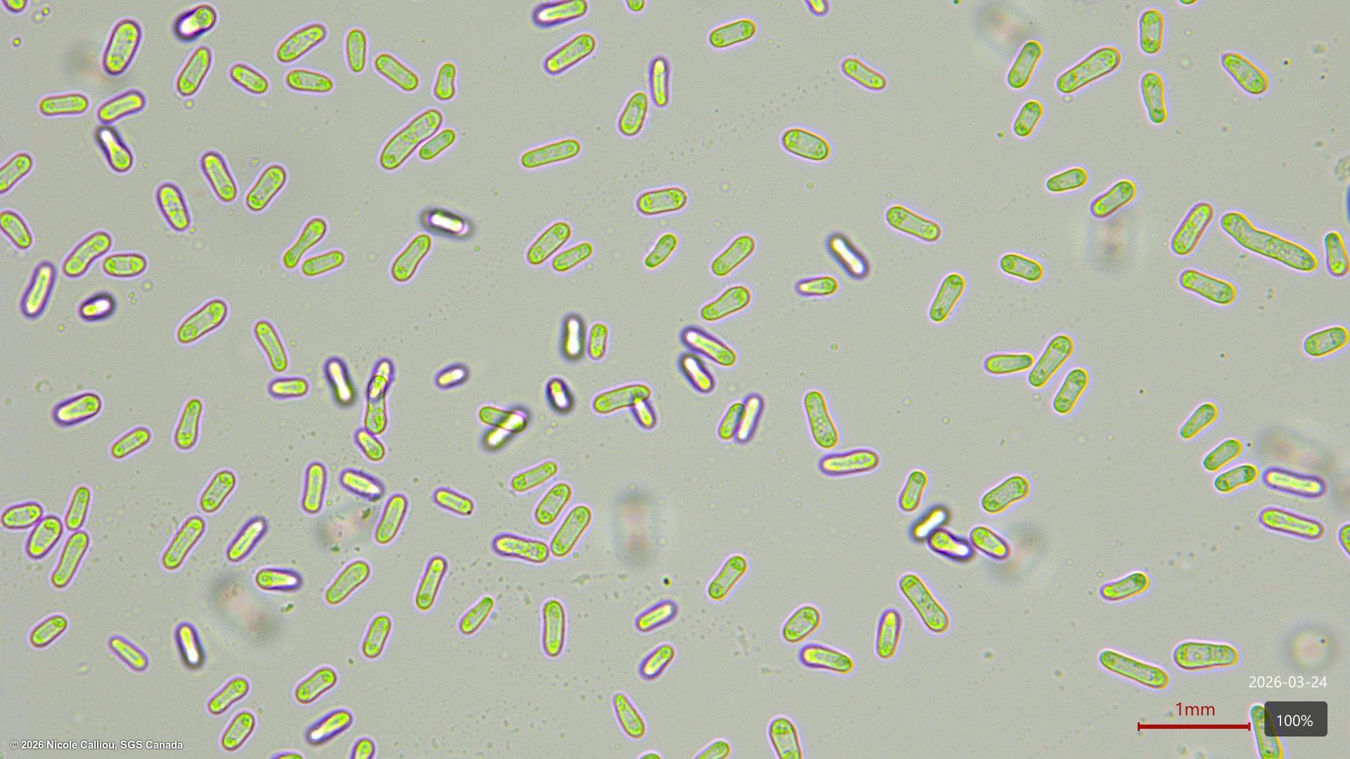
Ascochyta rabiei on Cicer arietinum
Image credits: Nicole Calliou, CA
On PDA, OR on MA, white to green colored mycelium develops slowly, development of pale brown to dark brown pycnidia occurs on surface or immersed. At 7 days, colony diameter varies between 10-35 mm on both media. Pycnidia usually release orange mucilaginous mass of conidia (cirrus). Doubtful colonies may be confirmed by the presence of pycnidia on seed coat or on media surface when examined at x25 magnification, using stereomicroscope. Pycnidia are 65-245 μm diameter, ostiole 30-50 μm wide. Further confirmation can be made by examining conidia present in pycnidia using compound microscope (x100-1000 magnification). Conidia are hyaline, straight, or slightly curved (0-)1 septate, some unicellular, slightly, or not constricted at the septum, rounded at each end, mostly 7-16 x 3-5 μm.
sterilisation: 1% NaOCl 4 minutes | growth time: 7 days | temperature: 22 ± 2 °C | lighting: Alternating 12 hours light and 12 hours dark | plate size: 90 mm | agar: PDA ( Neogen) |
DNA testing: N/A