Pathogen

Fusarium poae

Overview

Scientific name

Fusarium poae

Genus

Fusarium

EPPO code

FUSAPO

Common name

Root rot of grasses

Synonyms

Sporotrichum poae

Description

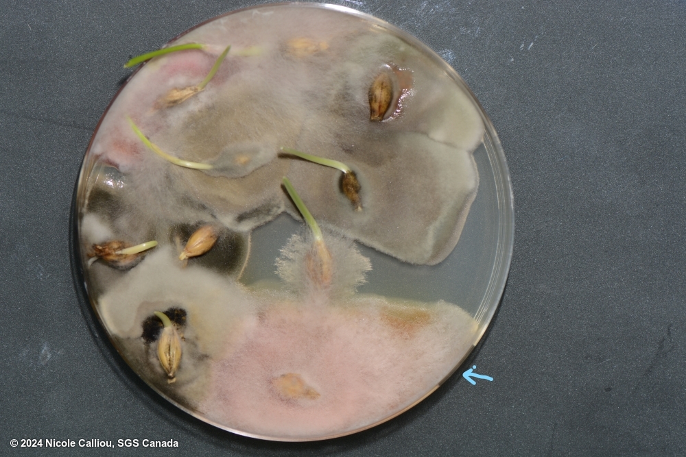
On PDA, growth is very rapid, with dense aerial, cotton like mycelium, often described as hairy or felted, and changing to powdery as microconidia are formed. Mycelium is generally white in colour, but can be yellow or pale pink, often becoming darker with age. Spores are readily formed in the mycelium. The under surface is usually pink to red, but colour variation including yellows can be observed. Sporodochia are rare.

On malt agar, the mycelium is dense, aerial, white to pink. Sporodochia are rare. 

Regardless of agar type, colonies will produce a very distinct scent, generally described as sweet. Often compared to apples or strawberries.

Conidiophores: Unbranched and branched monophialides. Microconidiophores are short and fat, almost globose, and quite distinctive. 

Macroconidia are generally rare, typically sickle shaped, 3-5 septate, but usually 3-septate. They will have a foot shaped basal cell. 

Microconidia are extremely abundant, globose, or oval in shape (napiform), sometimes with a distinct papilla, and usually 0- but sometimes 1-septate.

Chlamydospores occur infrequently and may be in clumps or chains. 

Images of Fusarium poae

Fusarium poae on Hordeum vulgare subsp. vulgare
click for more information

Fusarium poae on Hordeum vulgare subsp. vulgare

Image credits: Nicole Calliou, CA

Plates show both an F. poae (marked with blue arrow) and an F. graminearum colony. Spore image shows abundant microconidia, and microconidia on monophialide


sterilisation: 1% NaOCl 4 min | growth time: 5 days | temperature: 25+/-2C | lighting: 3:1 (fluorescent:nUV), within 60cm of light. Alternating light/dark in 12 hr intervals | plate size: 100mm | agar: PDA ( Neogen) |
DNA testing: None
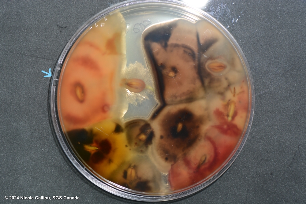
Fusarium poae on Hordeum vulgare subsp. vulgare
click for more information

Fusarium poae on Hordeum vulgare subsp. vulgare

Image credits: Nicole Calliou, CA

Plates show both an F. poae (marked with blue arrow) and an F. graminearum colony. Spore image shows abundant microconidia, and microconidia on monophialide


sterilisation: 1% NaOCl 4 min | growth time: 5 days | temperature: 25+/-2C | lighting: 3:1 (fluorescent:nUV), within 60cm of light. Alternating light/dark in 12 hr intervals | plate size: 100mm | agar: PDA ( Neogen) |
DNA testing: None
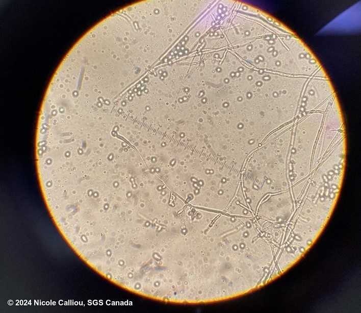
Fusarium poae on Hordeum vulgare subsp. vulgare
click for more information

Fusarium poae on Hordeum vulgare subsp. vulgare

Image credits: Nicole Calliou, CA

Plates show both an F. poae (marked with blue arrow) and an F. graminearum colony. Spore image shows abundant microconidia, and microconidia on monophialide


sterilisation: 1% NaOCl 4 min | growth time: 5 days | temperature: 25+/-2C | lighting: 3:1 (fluorescent:nUV), within 60cm of light. Alternating light/dark in 12 hr intervals | plate size: 100mm | agar: PDA ( Neogen) |
DNA testing: None