Fusarium langsethiae on Triticum spp.
Image credits: Nicole Calliou, NO
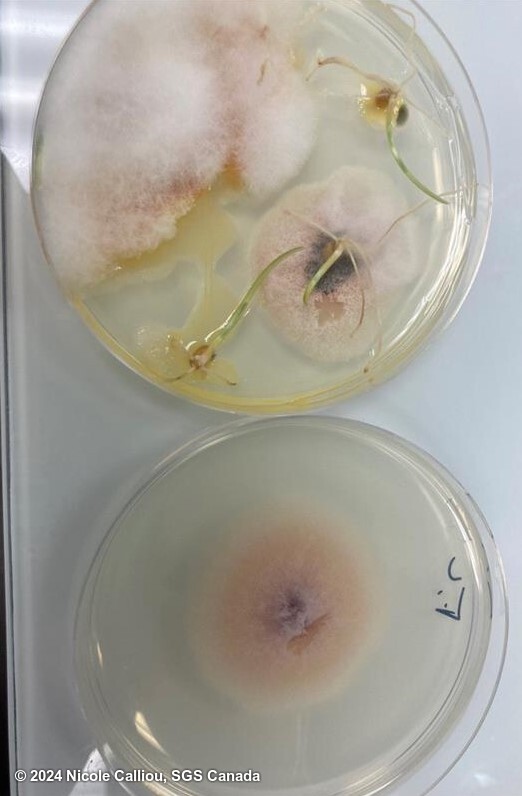
Top plate has a naturally infected wheat seed with typical sparse or powdery mycelium growth, whitish pink with a darker pink/purple center. Bottom plate shows the control culture. Only microconidia are produced, and typically in clumps. Macroconidia and chlamydospores are absent.
sterilisation: 1% NaOCl 10 min | growth time: 7 days | temperature: 21C | lighting: sunlight and darkness (12hr rotation) | plate size: 100mm | agar: PDA ( Neogen) |
DNA testing: None
Fusarium langsethiae on Triticum spp.
Image credits: Nicole Calliou, NO
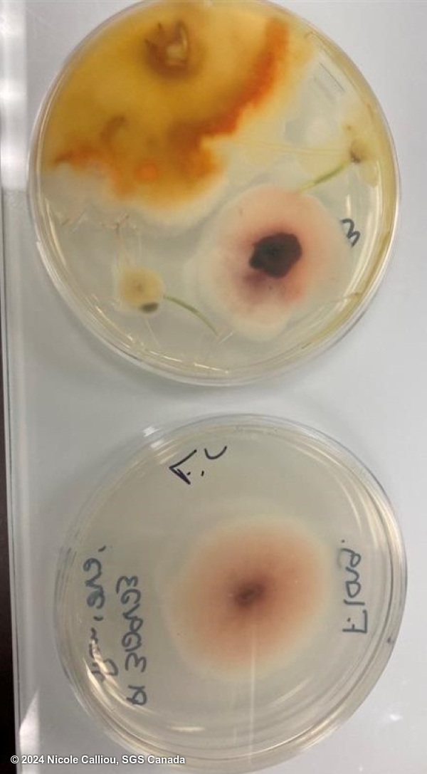
Top plate has a naturally infected wheat seed with typical sparse or powdery mycelium growth, whitish pink with a darker pink/purple center. Bottom plate shows the control culture. Only microconidia are produced, and typically in clumps. Macroconidia and chlamydospores are absent.
sterilisation: 1% NaOCl 10 min | growth time: 7 days | temperature: 21C | lighting: sunlight and darkness (12hr rotation) | plate size: 100mm | agar: PDA ( Neogen) |
DNA testing: None
Fusarium langsethiae on Triticum spp.
Image credits: Nicole Calliou, NO
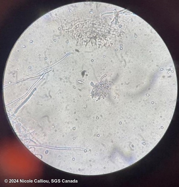
Top plate has a naturally infected wheat seed with typical sparse or powdery mycelium growth, whitish pink with a darker pink/purple center. Bottom plate shows the control culture. Only microconidia are produced, and typically in clumps. Macroconidia and chlamydospores are absent.
sterilisation: 1% NaOCl 10 min | growth time: 7 days | temperature: 21C | lighting: sunlight and darkness (12hr rotation) | plate size: 100mm | agar: PDA ( Neogen) |
DNA testing: None